
A fetus was discovered in the brain of a one year old child in a very unusual occurrence that was reported by doctors in Florida. After seeing symptoms like delayed development of motor skills, an increased head circumference, and accumulation of fluid in the brain, the pediatrician decided to send the child to the hospital.
After doing scans, the medical professionals discovered what they referred to as a “malformed monochorionic diamniotic twin” within the child’s skull. These twins come from the same fertilized egg and are identical, but in this case, one fetus had been enveloped by the other, creating what is known as “fetus in fetu” or “parasitic twin.”
Fetus in Fetu
“Fetus in fetu” is a rare phenomenon that happens in 1 in 500,000 live births. In most cases, the absorbed twin will cease its development while the other will keep growing. This will result in the absorbed twin appearing as a mass in the other fetus’s belly, which will be trapped behind the tissues that border the abdominal wall.
In this particular instance, the mass was found in the skull of the “host” fetus, and it most likely originated very early in the process of development, around the time when the fertilized egg is forming a cluster of cells known as a blastocyst.
Intracranial Fetus in Fetu
According to the report published in the journal Neurology, the “intracranial fetus-in-fetu is proposed to arise from unseparated blastocysts.” The cell clusters that were supposed to develop into two independent fetuses remained connected together.
The conjoined components eventually developed into the forebrain of the host fetus and encased the second embryo as the neural plate folded.
Just read about this and I’m stunned. Recently a 1-year-old girl with motor delay and enlarged head circumference was surgically operated for an intraventricular fetus-in-fetu and the post-op pictures are here: pic.twitter.com/vUvXIB6xXg
— Maahin (@dr_mxd) March 6, 2023
According to a 2020 case report in the journal World Neurosurgery, “Intracranial fetus in fetu is extremely rare with <20 reports published worldwide” The report also noted that the phenomenon usually occurs in the abdomen and is very rarely found in the head.
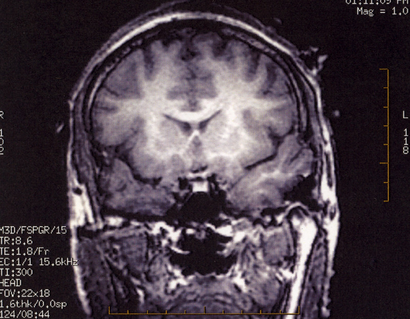
The brain scans of the one-year-old child indicated that the fetus included a vertebral column and two leg bones, the femur, and the tibia. The scans also revealed that the deformed baby had spina bifida, which is a disorder in which part of the spinal cord is revealed rather than being covered by the tissues of the back owing to a malfunction during development.
After being removed, it was revealed that the fetal mass had “upper limb and finger-like buds.”
Child’s Condition Following Surgery
The case report did not include details of the one-year-old’s condition following surgery, so it is unclear how the child has been affected by the surgery or the presence of the fetus in the brain.
This case highlights the importance of monitoring a child’s development and seeking medical attention if there are any signs of abnormal growth or development.
It also demonstrates the complexity of fetal development and the potential for rare anomalies to occur. Further research is required to understand the causes and potential treatments for “fetus in fetu” and similar anomalies.
The rarity of this case may make it a unique opportunity for medical professionals to learn more about the condition and potentially develop new treatments or therapies. As such, it may attract further interest and research in the medical community.
See all the latest news from Greece and the world at Greekreporter.com. Contact our newsroom to report an update or send your story, photos and videos. Follow GR on Google News and subscribe here to our daily email!